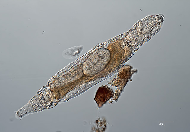
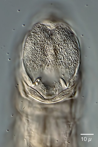
| Adineta editae, dorsoventral view, creeping; specimen from (1). |
|
|
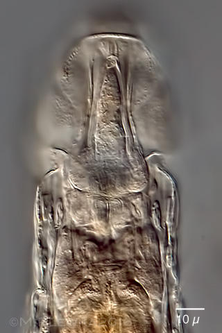
| Adineta editae, some aspects of the head; different focus planes. Upper left: dorsal view of the rostrum; probably showing brain and nerve cells. Upper left:optical transect showing the hypodermis cells and the semicircular-shaped rostrum. Middle left: focus plane on the ciliary field; middle right: focus plane on the two hook-shaped denticles of the rake apparatus. Lower left: ventral view of the ciliary field; the rostrum lamella is curled ventrally. lower right: ventral view of the rostrum lamella and cilia beneath the rostrum lamella. |
|
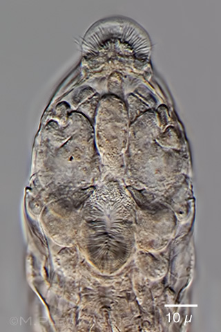
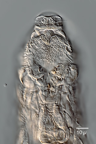
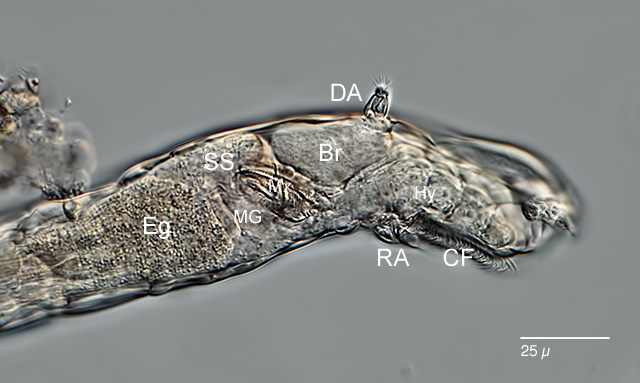
| Adineta editae, lateral view of the head region. Br: brain; CF: cilary field; DA: dorsal antenna; Eg: egg; Hy: hypodermis cells; MG: mastax ganglion; Mx: mastax; RA: rake apparatus; RG: rostral ganglion; SS. syncytial mass of the stomach. |
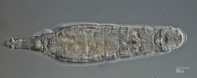 |
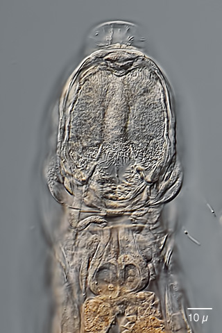
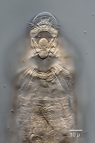
| Adineta editae, dorsoventral view of another specimen from (2) |
|
|
|
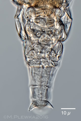
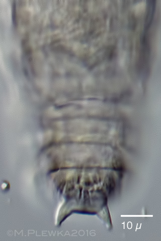
| Adineta editae, two aspects of the foot. Left image: foot of specimen from (1); right image shows the interspace between the spurs. |
|
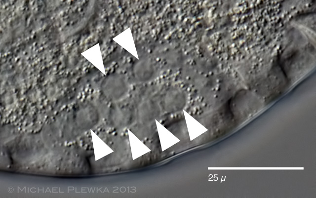 |
| Adineta editae, vitellarium of a slightly compressed specimen, showing at least 6 nuclei. If one assumes a closer relation between A. graciis and A. editae based on the similarity of the rake apparatus, then, on the other hand, there are differences with regards to the vitellarium, which has 4 nuclei in A. gracilis. |
|
| Location: Rocka Island, (Argentina archipelago) |
| Habitat: soil |
| Date: 17.01.2013 |
Samples provided by courtesy of Dr.N. Iakovenko (Ostrava; Czech Republic) and Dr. K. Janko (Libechov, Czech Republic).
Identification by courtesy of Dr. N.Iakovenko (Ostrava; Czech Republic). |